

 2 October, 2026
1. What Is an Accessory Cusp?
An accessory cusp (Dens evaginatus) is a term used to describe an abnormal anatomical shape of a tooth, characterized by a cusp-like or additional projection on the tooth surface. This condition commonly occurs on the inner surface of the upper teeth or on the occlusal surface of the lower premolars.
The accessory cusp has a structure similar to that of a normal tooth, consisting of enamel, dentin, and dental pulp. However, because the accessory cusp protrudes outward, the underlying pulp tissue may also extend into the cusp, forming thin pulp horns.
2. Why Does an Accessory Cusp Develop?
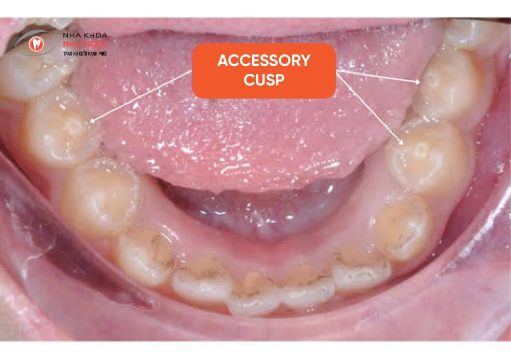
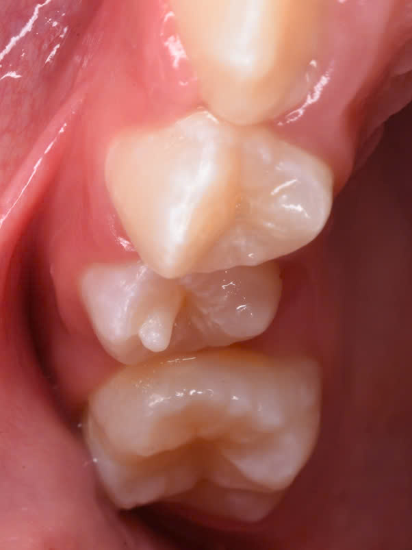
Image of a tooth with an accessory cusp on the occlusal surface
An accessory cusp results from abnormal tooth development during the morphodifferentiation stage, which occurs during the bell stage. It is characterized by abnormal proliferation of the inner enamel epithelium into the stellate reticulum of the enamel organ.
3. Potential Risks of an Accessory Cusp
An accessory cusp has a structure similar to that of a normal tooth, consisting of enamel, dentin, and dental pulp. However, because the cusp protrudes outward, the pulp tissue may also extend into it, forming thin pulp horns.
An accessory cusp can cause occlusal disturbances, resulting in interference during chewing and occlusal trauma. These may lead to complications that develop silently and are often difficult for patients to detect themselves, such as cervical resorption, gingival recession, dental caries, periapical abscesses, and pulp necrosis.
These abnormalities can be detected relatively easily by visual examination. Parents can simply observe their child’s teeth and, if an accessory cusp is noticed, take the child to a specialized medical or dental facility for timely evaluation and intervention.
4. Treatment of an Accessory Cusp
An accessory cusp may be treated by gradual reduction or complete removal of the cusp, followed by remineralization with fluoride varnish. However, cusp reduction must be performed carefully and depends on several factors, including whether the accessory cusp causes occlusal interference, the thickness of the enamel and dentin overlying the pulp horn, and the patient’s sensitivity. Therefore, dental X-rays should be taken to assess the thickness of the enamel and dentin over the pulp horn and determine an appropriate treatment plan.
- If the accessory cusp does not interfere with the occlusion, it should be protected with composite resin by covering the surrounding area with composite resin. This helps prevent dental caries caused by the anatomical shape of the accessory cusp, which may make oral hygiene more difficult. Such teeth should be monitored regularly every 6 months.
- If the accessory cusp causes occlusal interference, treatment may involve gradually reducing its height to allow the formation of reparative dentin over the pulp and remineralization of the enamel with fluoride varnish. The treatment steps include:
- Using a fine-grit diamond bur at high speed with water cooling.
- Performing the reduction over multiple appointments. The amount of reduction at each appointment depends on the thickness of the enamel and dentin layer overlying the pulp.
 |
 |
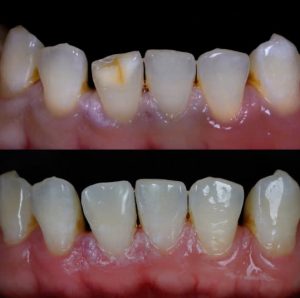
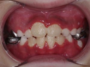
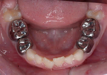
Before and After Images of the Tooth Following Accessory Cusp Removal
- The interval between two reduction appointments is approximately 3 months, with follow-up for at least 12 months.
- The height of the accessory cusp is reduced until there is no longer any occlusal interference when checked with articulating paper.
- If the reduction results in pulp exposure, direct pulp capping should be performed using a biocompatible material such as MTA or Biodentine, followed by continued monitoring.
In cases where a permanent tooth with an immature apex develops pulp necrosis due to an accessory cusp, the indicated treatment is regenerative endodontic therapy using a dental operating microscope and biocompatible materials such as Biodentine or MTA. This approach helps restore pulp-related function and support continued root development and apical closure, allowing the root to develop optimally.
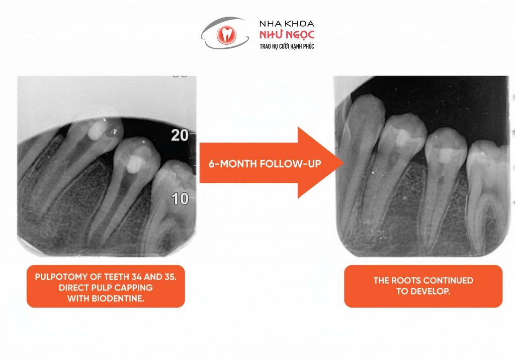
This technique requires dentists with advanced expertise in endodontic treatment and the use of a dental operating microscope to precisely control and manage the pulp tissue, thereby helping achieve optimal treatment outcomes for patients.